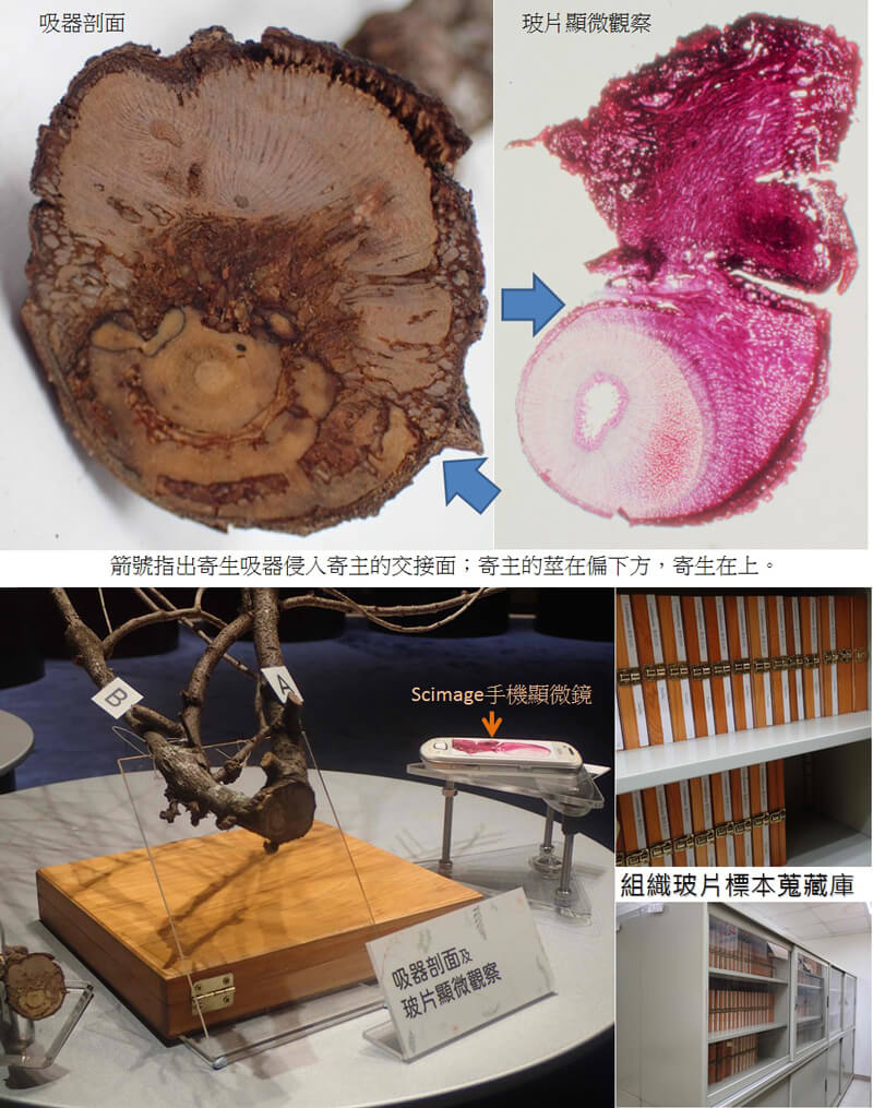
Cross-section of haustorium and microscopic observation of glass slides
Looking at a cross-section of the haustorium, the integration of tissues is seamless, meaning that it can easily and seamlessly penetrate and obtain water and nutrients from the host. A denotes the parasite, B denotes the host.
The arrowhead indicates the point where the haustorium of the parasite invades the host. In the center of the cross-section of the host’s trunk is the pith, which is surrounded by xylem. The parasite’s haustorium penetrates the surface layer, cortex, and phloem. It can go as deep as the newly produced secondary xylem.
This museum’s curators carry out research on and prepare wood samples and tissue slides, according to the strict international herbarium and wood collection standards, with the mission to preserve natural objects.
Use the Scimage smartphone microscope to observe slides. The cross section of the host’s trunk is arch-shaped. The upper part is the longitudinal section of the parasite’s stem. The haustorium resembles an anchor that is embedded in the host’s trunk. In this example, there is invasion of tissue that connects to the xylem of the host. The ploem and cortex, outside the xylem, are also in the occupied area. This means that the parasite invades the host through the haustorium and directly obtains water and nutrients from the host.
For an introduction to the Scimage smartphone microscope, refer to the article entitled “In Fashion Microscopic Observation” in issue 354 of the NMNS newsletter: https://libknowledge.nmns.edu.tw/nmns/lib/PDF/106/354/4.pdf
(in Chinese).
You can also go to NMNS’ Naturalist Center to view or use the Scimage smartphone microscope.